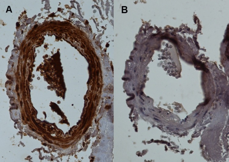
Fig 10. Immunohistochemistry for human artery with antibody against GJC3, hCx30.2/31.3.
(A) H-86—sc-68376, Santa Cruz Biotechnology, Inc., vs. (B) control section, both micrographs at 20 x magnification. Brown staining in (A) indicates selective localisation of GJC3 in media and endothelium of the artery.

