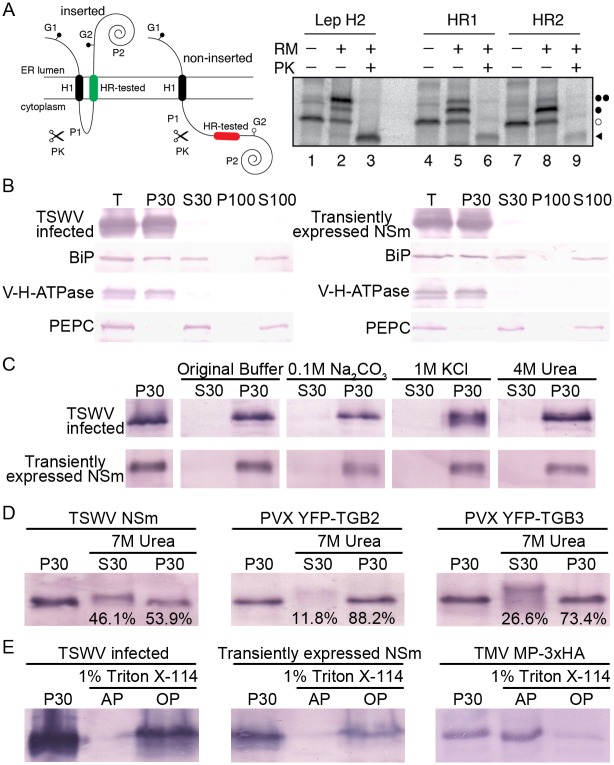
Fig 1. The NSm protein of tomato spotted wilt tospovirus (TSWV) is physically associated with cellular membranes.
(A) Insertion assay of NSm hydrophobic regions 1 (HR1) and 2 (HR2) into microsomal membranes using the Lep’ construct. Schematic representation of the Lep-derived construct (Lep’) is shown in upper panel. In this Lep’ construct, H1, derived from the glycosylation acceptor site (G2) at the beginning of the P2 domain, will be modified only if the tested HR inserts into the membrane; the G1 site, embedded in an extended N-terminal sequence of 24 amino acids, is always glycosylated. Results of in vitro translation and membrane insertion experiments are shown in the lower panel. Bands of nonglycosylated protein are indicated by a white dot; singly and doubly glycosylated proteins are indicated by one and two black dots, respectively. The protected glycosylated HRs/P2 fragment is indicated by a black triangle. (B) Association of NSm with membrane factions. Total lysate (T) from TSWV-infected or NSm expressing leaves were fractionated into 30,000 × g pellet (P30), 30,000×g supernatant (S30), 100,000×g pellet (P100) and 100,000×g supernatant (S100), and analyzed by immunoblots using antibodies against NSm. The vacuolar H-ATPase (V-H-ATPase) subunit E, phosphoenolpyruvate carboxylase (PEPC) and the luminal binding protein (BiP) were used as a microsomal marker, soluble marker and ER marker, respectively, in the fractionation analysis. (C) Biochemical characterization of NSm associated with membranes. The P30 pellet fraction was treated with original lysis buffer, 0.1 M Na2CO3, 1 M KCl, or 4 M urea, respectively, then separated into supernatant (S30) and pellet (P30) fractions and analyzed by immunoblots using anti-NSm antibodies. (D) Membrane association analysis of TSWV NSm, PVX TGB2 and TGB3 after treatment with 7 M urea. The percentage of proteins eluted in the S30 supernatant or remaining in the P30 pellet, are given at the bottom of the corresponding lanes. (E) Triton X-114 partitioning analysis of TSWV NSm and TMV MP. P30 pellet, aqueous phases (AP) and organic phases (OP) were analyzed by immunoblotting using anti-NSm and anti-HA antibodies, respectively.

