
Fig 2. TSWV NSm is localized with the ER and plasmodesmata (PD).
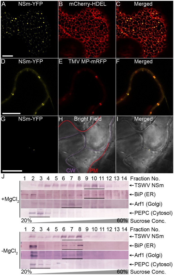
(A-C) Colocalization of NSm-YFP with the ER labeled by mCherry-HDEL at 28 h post infiltration (hpi). Bar, 10 μm. (D-F) Colocalization of NSm-YFP with PD labeled by TMV MP-mRFP at 28 hpi. Bar, 10 μm. Different single planes were used to focus on the ER membrane in panels A-C panels and on the periphery of the cell in panels D-F. (G-I) Plasmolysis assay for PD localization of NSm. N. benthamiana leaves were agroinfiltrated with NSm-YFP, then infiltrated with 10% NaCl at 28 h post agroinfiltration; plasmolyzed cells in the leaf were immediately examined using CLMS. The cell wall (CW) and cytoplasmic membrane (PM) after plasmolysis are marked, respectively, by a purple line and red line. (J) Cofractionation of NSm protein with ER. Extracts of plants transiently expressing NSm were centrifuged on a 20–60% sucrose gradient in the presence or absence of MgCl2. Fractions from top to bottom (1 to 14) were analyzed by immunoblots using anti-NSm, anti-BiP, anti-Arf1 and anti-PEPC antibodies to detect NSm, ER luminal protein, Golgi and soluble protein, respectively.