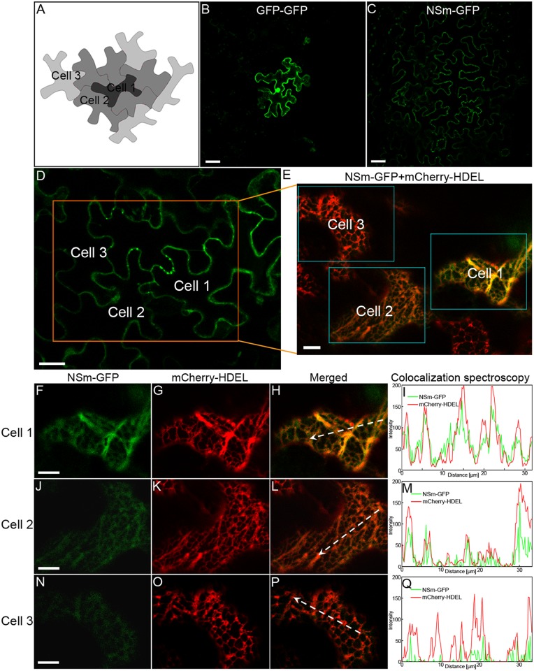
Fig 3. Localization of NSm-GFP in the ER membrane network in bombarded cells with the fusion protein and in neighboring cells receiving the fusion protein in leaf epidermis of N. benthamiana.
(A) Scheme of cell-to-cell transport of NSm in leaf epidermis of N. benthamiana by bombardment. (B and C) Cell-to-cell movement of GFP-GFP (B) and NSm-GFP (C) in leaf epidermis of N. benthamiana. Bar, 50 μm. (D-Q) NSm-GFP was localized in the interconnected ER network in cells bombarded with the fusion protein and in neighboring cells that subsequently received the protein. A low magnification image to show that NSm moved intercellularly after bombardment (D). Bar, 20 μm. A region with three cells showing NSm movement (Cell 1 to Cell 3) in image D was magnified (boxed region) to show colocalization of NSm-GFP with the ER labeled by mCherry-HDEL (E). Bar, 10 μm. The boxed region in image E corresponding to the respective initially bombarded cell (Cell 1) and the second (Cell 2) and third layer (Cell 3) of cells into which NSm moved was further split into three channels to show colocalization of NSm-GFP with the ER labeled by mCherry-HDEL (Cell 1, F-H; Cell 2, J-L; Cell 3, N-P). Colocalization of NSm and ER in the respective cells was further analyzed by overlapping fluorescence spectra (I, M and Q). Bar, 10 μm.

