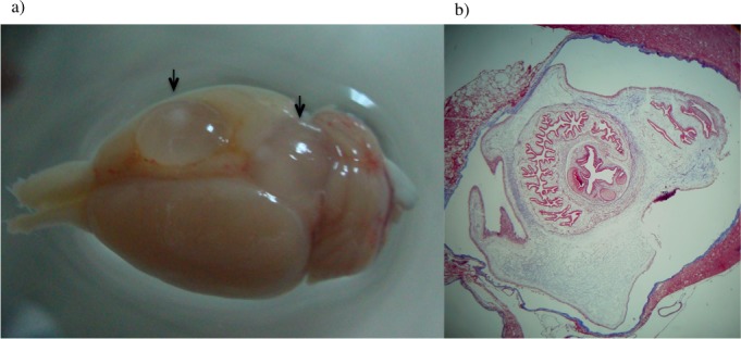
Fig 6. Rat brain infected with T. solium postoncospheral forms.

(a) Rat brain 4 months post infection showing two extraparenchymal cysticerci (arrows). (b) Coronal sections of the brain containing an intraparenchymal cysticercus stained with Masson’s Trichrome staining showing tegument and scolex.
