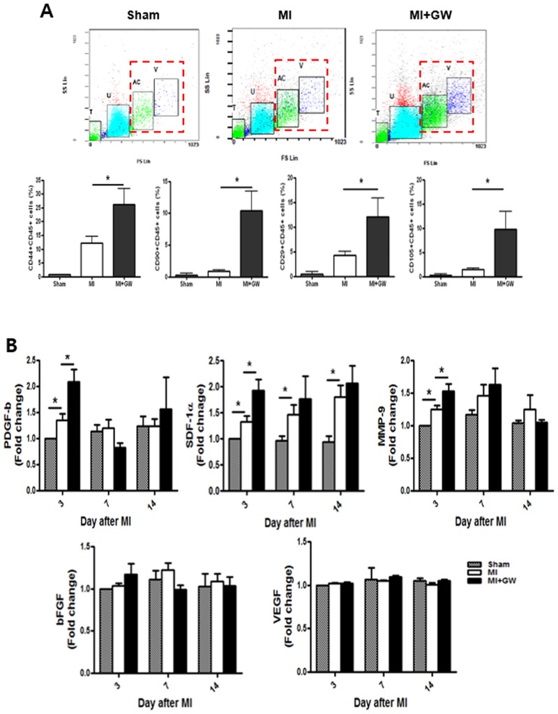
Fig 6. Effects of GW610742 on MSC migration into the infarcted hearts.
(A) Rat cardiac cells on days 3 after MI were prepared from the ventricles, and adherent cells were stained with each antibody. “T” and “U”-gated cells (floating cells also have “T” and “U”-gated cells) were ruled out for analysis, and “AC” and “V”-gated cells were verified for MSC-phenotype analysis. MSC-positive cells were counted in “AC” and “V”-gated cells. (B) Serum from each group on each day was measured for PDGF-b, SDF-1α, MMP-9, bFGF, and VEGF using specific ELISA kit. Values are represented as the mean ± SEM. *P <0.05. MSC, mesenchymal stem cell; MI, myocardial infarction; PDGF-b, platelet-derived growth factor subunit B; MMP-9, matrix metallopeptidase 9; SDF-1α, stromal-derived factor-1 alpha; bFGF, basic fibroblast growth factor; VEGF, vascular endothelial growth factor. Sham (n = 3), MI (n = 7), MI + GW (n = 7).

