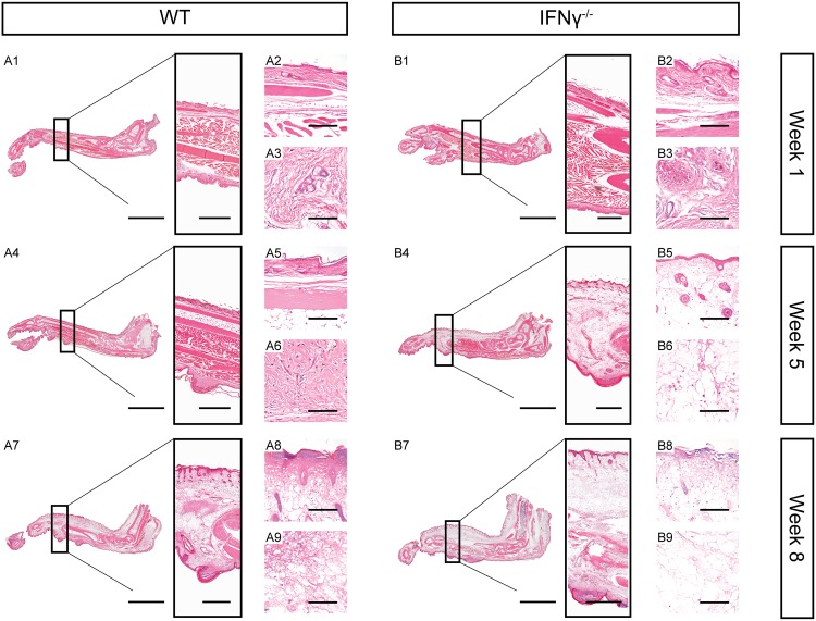
Fig 2. Extensive tissue necrosis and oedema formation in mice lacking IFNγ.
HE stained histologic sections of foot pads from representative WT (A) and IFNγ-/- (B) mice 1, 5 and 8 weeks after infection with M. ulcerans. Scale bars represent 5 mm (A1, A4, A7, B1, B4 and B7, left), 1 mm (A1, A4, A7, B1, B4 and B7, box), 150 μm (A2, A5, A8, B2, B5 and B8) and 80 μm (A3, A6, A9, B3, B6 and B9).

