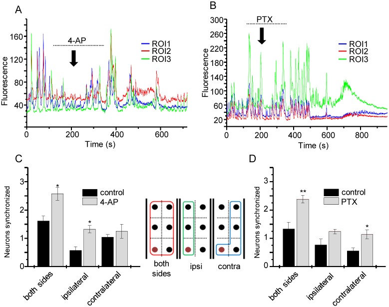
Fig 3. Proconvulsants increase synchronicity of neuron activity.
A) GCaMP5 fluorescence from single aCC neurons from consecutive segments show hyperactive and synchronised pattern changes following 4 min application of 4-AP (3 mM). Dotted line shows period when 4-AP was perfused and downward arrow indicates time 4-AP entered the bath. The time difference results from the length of tubing between gravity-driven perfusion system and the recording chamber. B) PTX (5μM) perfusion similarly increases neuron activity. Dotted line shows period when PTX was perfused and downward arrow indicates time PTX entered the bath. C, D) Analysis of number of synchronised aCC cells before and after 4-AP (C) and PTX (D) application (values are mean ± sem for n = 6 and 5, respectively). Synchrony was compared between a fixed ROI (labelled brown in the inset) and compared to all ROIs (red box on inset) and with those on the ipsilateral (green box) or contralateral (blue box) sides.

