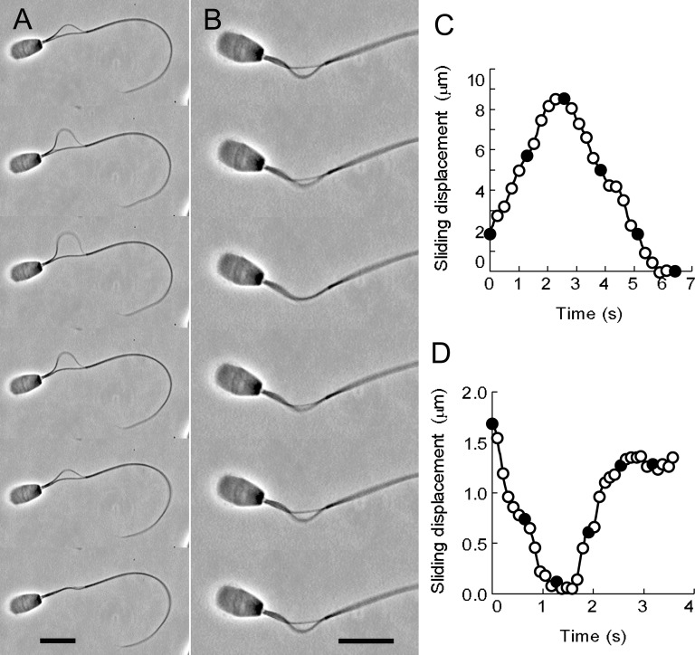
Fig 1. The oscillatory sliding movement of the doublet microtubules of bull sperm models.
(A) Phase-contrast video micrographs showing the oscillatory sliding movement of the doublet microtubules extruded by synchronous sliding. (B) Phase-contrast video micrographs showing the oscillatory sliding movement by metachronal sliding. The free-Ca2+ solution concentration was adjusted to 10−9 M. Time interval between successive images is 1.28 s in (A) and 0.64 s in (B). The profiles of the microtubule sliding displacement of A and B are shown in (C) and (D), respectively. The sliding velocity of the synchronous sliding movement was 3.02 μm/s, which was calculated between frames 1 and 2 in A (C). The sliding velocity of the metachronal sliding movement was 1.23 μm/s between frames 1 and 3 in B (D). Filled circles in C and D are the values of the sliding displacement obtained from the frames shown in A and B. Bars = 10 μm.

