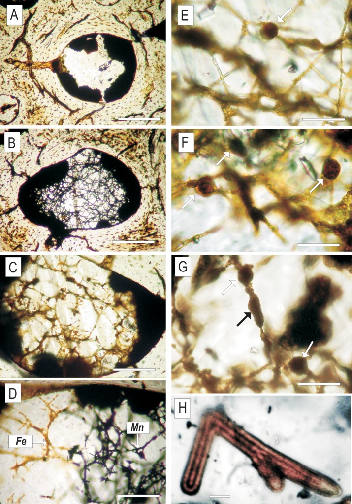
Fig 2. Transmitted light images of mycelia preserved inside dinosaur bone.
(A‒C) Bifurcating translucent hyphae embedded in calcite inside the marrow cavity of ZPAL MgD I/alt bone (D) A network of hyphae embedded in calcite filling a bone void; note the changing mode of hyphae mineralisation, from black manganese oxides to brown iron oxides (E‒G) Network of hyphae with putative asexual reproductive bodies (?spores) indicated with arrows (H) Fragment of iron-manganese-oxide-permineralised mycelium showing the distinctly siphonous organisation of the branching hyphae. Scale bars: (A‒C) 100μm (D) 50 μm (E-G) 20 μm (H 5) μm.

