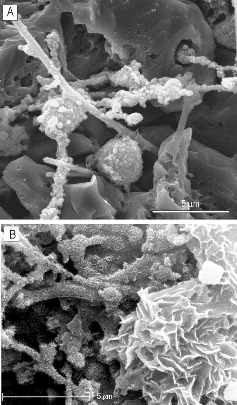
Fig 5. SEM images of a mineralised mycelium exposed with formic acid etching from polished thin section ZPAL MgD I/8 bone.
(A) Hyphae and putative asexual reproductive bodies (?spores) covered with Fe/Mn nanograins (B) A fragment of organically preserved hyphae (left) and a rosette-flower-like pattern of ferromanganese micronodules precipitated into the mycelium biomass (right).

