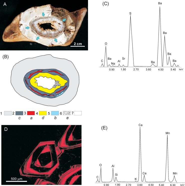
Fig 8. Successively precipitated mineral phases in marrow cavity.
(A) Cross section of a femur bone (specimen ZPAL MgD I/8) showing a marrow cavity (delimited by arrows) filled almost entirely with largely microbially-mediated mineral succession (B) Schematic illustration of the mineral succession imaged in (A); 1 –massive bone, 2 –micritic calcite filling the marrow cavity colonised by fungi, 3 –mineralised fungal biofilm, 4 –abiotically precipitated crystalline calcite, 5 –barite, 6 –intrusion of clastic sediment, 7 –open space of the marrow cavity; lowercase letters indicate the sequence of precipitation of mineral phases from (a) to (e) (C) SEM/EDS spectrum documenting the presence of barite, precipitated in the marrow cavity as indicated in the mineral succession diagram in (B) (D) Cathodoluminescence image of banded calcite representing the later stage of mycelia mineralisation after the earlier diagenetically precipitated ferromanganese. (E) SEM/EDS spectrum documenting the presence of calcite.

