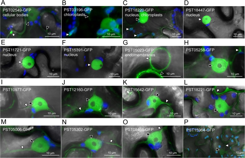
Fig 1. Seven candidate effectors show specific accumulation patterns in leaf cells.
Live-cell imaging of the 16 candidate effector-GFP fusion proteins accumulating in distinct subcellular compartments of N. benthamiana leaf cells. Proteins were transiently expressed in N. benthamiana leaf cells by agroinfiltration. Live-cell imaging was performed with a laser-scanning confocal microscope two days after infiltration. GFP and chlorophyll were excited at 488 nm. GFP (green) and chlorophyll (blue) fluorescence were collected at 505–525 nm and 680–700 nm, respectively. Images are single optical sections of 0.8 μm or a maximal projection of up to 47 optical sections (max. z-stack of 37.6 μm). Images displayed are overlays of the GFP signal, the chlorophyll signal, and bright field. For A-G, specific cellular compartments in which the GFP signal accumulates are indicated. White arrowheads indicate GFP-labelled cytosolic bodies (A), chloroplasts (B-C), nuclei (D-F), nuclear surrounding (G), or cytosolic fractions (H-P). Black arrowheads indicate GFP-labelled small cytosolic bodies (A), a stromule (B), a nucleus (C), the plasma membrane (G), or nuclei (H-P). In (P), the low level of accumulation of the fusion protein imposed higher laser power and gain, which resulted in non-specific signal for the GFP channel being visible in chloroplasts and ostiole edges.

