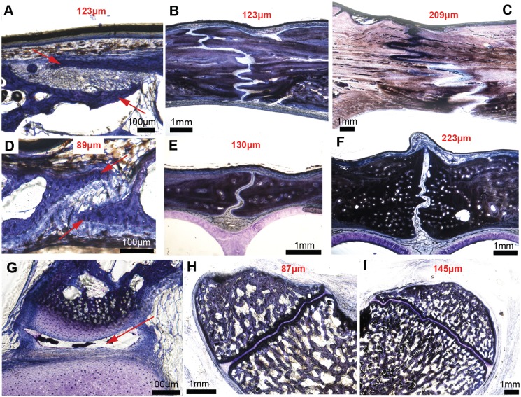
Fig 8. Transverse sections through the frontoparietal suture, the internasal suture and the basioccipital-exoccipital synchondrosis in an ontogenetic series of three American alligator heads, stained with Toluidine-blue.
A-C, Frontoparietal suture; D-F, internasal suture; G-H, Basioccipital-exoccipital synchondrosis. The youngest specimen MOR OST 1647 is a few days old and is shown in the first column (A, D, G); the second column shows a 4 to 5 year-old specimen MOR OST 1797 (B, E, H); the third column shows a 9 to 12 year-old sexually mature specimen, MOR OST 1798 (C, F, I). Sutures of the youngest specimen show two thin bone struts overlapping each other (A, D, red arrows). The basioccipital-exoccipital synchondrosis (G) in this same specimen is not fully formed (and appears to have a synovial cavity, red arrow). Interdigitations increase drastically in the frontoparietal suture, but only slightly in the internasal suture (being a midline suture) and in the basioccipital-exoccipital synchondrosis. These sections do not show any sutural bony fusion. The average sutural and synchondroseal widths are marked in red on each photomicrograph, and it is clear that these widths increase as ontogeny progresses.

