. 2015 Dec 11;4:e09003. doi: 10.7554/eLife.09003

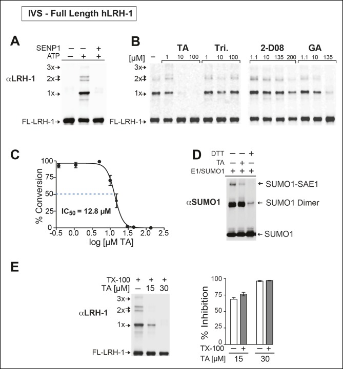
Figure 4. TA is a detergent-resistant inhibitor of substrate sumoylation in vitro.

(A) In vitro sumoylation (IVS) of recombinant full length (FL)-hLRH-1, without ATP, with ATP or with ATP and recombinant SENP1 added to IVS reactions as described in ‘Materials and methods’. (B) IVS assays with increasing tannic acid (TA), trifluidine (Tri.), 2-D08, and ginkgolic acid (GA). Sumoylated and unsumoylated FL-hLRH-1 are indicated with arrows as detected with anti-LRH-1 antibody. (C) IC50 of TA in FL-hLRH-1 IVS assay. Data are represented as mean ± SEM from at least three independent replicates. (D) Formation of E1 thioester with or without TA, in non-reducing conditions (-DTT). Effects of TA (10 µM) on formation of SUMO-E1 complex (SUMO-SAE1, top band) compared to reducing conditions without TA (-DTT, last lane). SUMO1 dimers are formed in non-reducing conditions (SUMO1 Dimer). E1 thioester formation assays are initiated by addition of freshly prepared ATP (10 mM) and described in ‘Materials and methods’. Anti-SUMO1 antibody was used to detect SUMO1 species. (E) Levels of sumoylated and unsumoylated FL-LRH-1 in IVS assay with TA (15 and 30 μM) and in the presence or absence of Triton X-100 in vitro (left panel). Bar graph of quantified data showing percent inhibition of hLRH-1 sumoylation by TA and with or without Triton X-100 (right panel).

DOI: http://dx.doi.org/10.7554/eLife.09003.012

Figure 4.

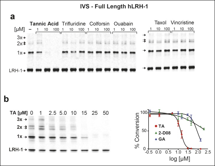
Figure 4—figure supplement 1. Effects of TA, other candidate hits, and published sumoylation inhibitors in an IVS assay of full-length hLRH1.

Figure 4—figure supplement 1.

(A) Dose-dependent inhibition of full-length (FL)-hLRH-1 by TA compared to other top candidate hits from primary screen as assayed by IVS. IVS assay and immunoblotting conditions used to detect hLRH-1 species are described in Materials and Methods. Sumoylated hLRH-1 (1x, 2x, 3x) and unmodified LRH-1 (LRH-1) species are indicated by arrows. (B) IVS assays of FL-hLRH-1 were performed with increasing concentrations of TA are shown (left panel) and plotted as normalized values in graph (right panel). Effects of two other published sumoylation inhibitors, 2-D08 and GA are also shown in graph. IVS data was normalized to DMSO control for each compound, and then plotted as percent conversion per log10 [µM] concentration. Curve fitting of data is described in ‘Materials and methods’. GA: Ginkgolic acid; IVS: In vitro sumoylation; TA: Tannic acid.
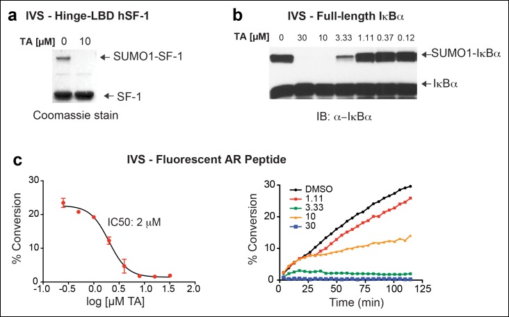
Figure 4—figure supplement 2. IVS of multiple substrates inhibited by TA.

Figure 4—figure supplement 2.

(A) Coomassie staining of recombinant hinge-LBD mSF-1 protein (aa178-462) with DMSO (0) or with 10 µM TA. This protein fragment of mSF-1 contains only one of the two conserved sumoylation consensus sites at K194. Migration of SUMO1-SF-1 and unmodified SF-1 hinge-LBD (SF-1) are indicated by arrows. (B) IVS of full length IκBα without (0) or with increasing concentrations of TA, as indicated, with SUMO-IκBα and unmodified IκBα indicated by arrows. (C) Dose-dependent inhibition of IVS of fluorescently labeled AR peptide by TA with the IC50 provided. Data are plotted as percent conversion versus TA concentration (log10 [µM], left panel). Real-time sumoylation of AR peptide (% Conversion) are plotted at different concentrations of TA (right panel). IVS conditions and detection of AR peptide sumoylation by electrophoretic mobility shift assay are previously described in Kim et al., (2013).
IVS: In vitro sumoylation; TA: Tannic acid.