
Figure 2.
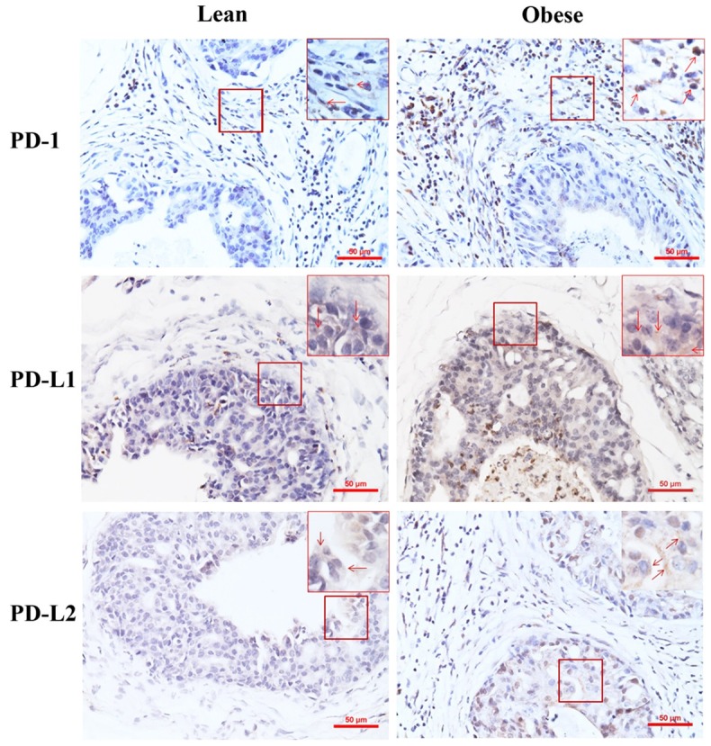
Representative photomicrographs of immunohistochemical staining of lean and obese mouse prostate tumors. Original magnification, x200. In highlighted frames, original magnification, x400; arrows indicate the positively stained cells.
Official websites use .gov
A
.gov website belongs to an official
government organization in the United States.
Secure .gov websites use HTTPS
A lock (
) or https:// means you've safely
connected to the .gov website. Share sensitive
information only on official, secure websites.
Representative photomicrographs of immunohistochemical staining of lean and obese mouse prostate tumors. Original magnification, x200. In highlighted frames, original magnification, x400; arrows indicate the positively stained cells.