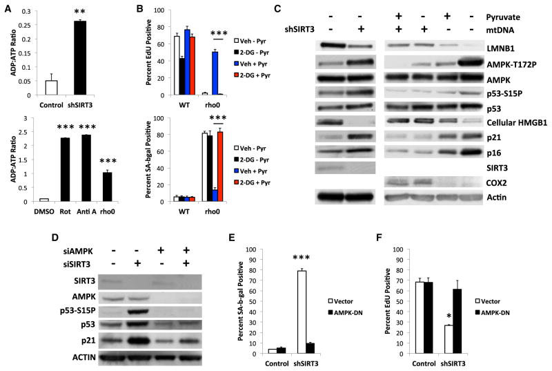
Figure 4. MiDAS Depends on AMPK Activation.
(A) Cells infected with control or shSIRT3-expressing lentiviruses (upper panel), or treated with DMSO, Rot, Anti A, or ethidium bromide (EtBr) (lower panel) for 10 days were analyzed for ADP:ATP ratios.
(B) WT or rho0 cells cultured + or −pyruvate and/or 2-DG for 10 days were analyzed for EdU labeling (upper panel) or SA-Bgal (lower panel).
(C) Control or shSIRT3-expressing cells were cultured for 10 days after infection. WT (mtDNA+) and rho0 (mtDNA−) cells were cultured + or − 1 mM pyruvate for 10 days, lysed, and analyzed by immunoblotting for total and phosphorylated AMPK and p53 and intracellular LMNB1, HMGB1, p21WAF1, p16INK4a, SIRT3, COX2, and actin.
(D) Control (siSIRT3/AMPK−), SIRT3 (siSIRT3+), and AMPKα-1/2 (siAMPK+) siRNAs were transfected into cells, which were analyzed by immunoblotting 4 days later for the indicated proteins.
(E and F) Cells infected with control or dominant-negative AMPK (AMPK-DN)-expressing lentiviruses, then control or shSIRT3-expressing lentiviruses were analyzed 7 days later for (E) SA-Bgal and (F) EdU incorporation. Bar graphs indicate mean + SEM.

