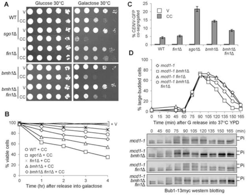
Figure 3.

The premature SAC silencing in bmh1Δ mutants is partially Fin1-dependent. (A) Cells were 10-fold diluted and spotted onto glucose and galactose plates. The plates were incubated at 30°C for 2 days before scanning. (B) fin1Δ deletion partially suppresses the viability loss of bmh1Δ cells overexpressing CIK1-CC. Asynchronous cells with either a vector or a PGALCIK1- CC plasmid were grown to log-phase in raffinose medium and then galactose was added to a final concentration of 2%. The cells were collected over time and spread onto YPD plates to determine the plating efficiency after overnight incubation. (C) fin1Δ deletion partially suppresses chromosome mis-segregation in bmh1Δ mutant cells. G1-arrested CEN4-GFP TUB1-mCherry cells with indicated genotypes in raffinose medium were released into galactose medium for 120 min at 30°C. The cells were collected to visualize the spindle morphology and CEN4-GFP distribution. The percentage of cells with co-segregated CEN4-GFP was counted (n > 100). The percentage is the average from three independent experiments. (D) fin1Δ deletion partially suppresses the premature Bub1 dephosphorylation in bmh1Δ cells lacking tension. BUB1-13myc cells with the indicated genotypes were synchronized in G1 phase and then released into YPD at 37°C to inactivate cohesin Mcd1. The cells were collected every 15 min for the budding index and the examination of Bub1 phosphorylation.
