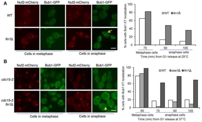
Figure 7.

Fin1 promotes the dissociation of Bub1 from kinetochores in anaphase. (A) The kinetochore localization of Bub1 in WT and fin1Δ mutant cells. Cells were synchronized in G1 and then released into cell cycle at 25°C. The localization of Bub1-GFP and a kinetochore protein Nuf2-mCherry was examined over time. At 75 min after release, the majority of cells are in metaphase, and the percentage of metaphase cells with kinetochore-localized Bub1 is shown. The percentage of anaphase cells that show kinetochore-localized Bub1 was counted for the 90 and 105 min samples. Some representative cells are shown in the left panel. Scale bar, 5μm. (B) The kinetochore localization of Bub1 in cdc15-2 mutants. G1-arrested cdc15-2, cdc15-2 fin1Δ and cdc15-2 ase1Δ cells with Nuf2-mCherry Bub1-GFP were released into 37°C YPD. The percentage of metaphase cells with kinetochore-localized Bub1 was counted at 60 min as majority of the cells are in metaphase. The percentage of anaphase cells with kinetochore-localized Bub1 was assessed at 75, 90 and 105 min. Some representative cells are shown in the left panel. Scale bar, 5μm.
