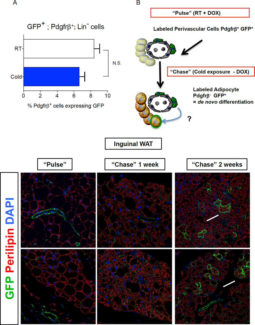
Figure 4. Pdgfrβ+ mural cells contribute to beige adipocyte formation in adult WAT after prolonged cold exposure.
(A) Percentage of Pdgfrβ+;Lin− cells expressing GFP (GFP+; Pdgfrβ+; Lin−) in WAT depots of Zfp423GFPB6 male mice housed at room temperature or cold exposed (6°C) for 2 weeks. Bars represent mean ± SEM. n= 5–6 animals for each group. (B) “Pulse-Chase” lineage tracing approach. DOX-containing food (600 mg/kg) is administered to MuralChaser mice for 9 days at room temperature to label Pdgfrβ+ cells (“Pulse”). Afterwards, animals were immediately harvested (Pulse) or are housed in the cold (6 °C) for either 1 or 2 weeks in the absence of DOX (Chase). mGFP+ multilocular adipocytes represent de novo differentiated fat cells formed during the cold exposure period. (C–D) Confocal immunofluorescence image of inguinal WAT from MuralChaser mice dox-treated for 9 days. Parrafin–embedded sections are immunostained with antibodies recognizing mGFP and perilipin. mGFP staining is confined to the vasculature at this stage. (E–F) Confocal immunofluorescence image of inguinal WAT from MuralChaser mice after 1 week of cold exposure (after removal of DOX). mGFP staining is still largely confined to the vasculature at this stage. (G–H) Confocal immunofluorescence image of inguinal WAT from MuralChaser mice after 2 weeks of cold exposure (after removal of DOX). Several small clusters of mGFP+ multilocular adipocytes can now be observed, indicating a contribution of Pdgfrβ+ mural cells to beige adipogenesis during prolonged cold exposure. All images photographed at 63× magnification.

