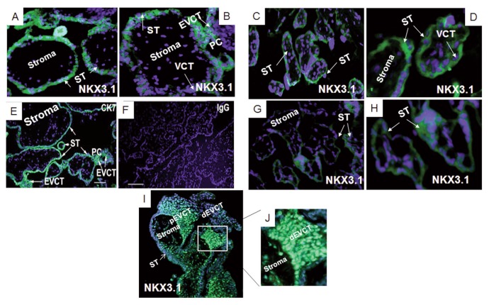
Figure 3.
NKX3.1 protein expression in first and third-trimester placental sections. Localization of NKX3.1 protein in a first-trimester placenta (A and B, n = 6) in a third-trimester FGR placenta (G and H, n = 6) and a gestation-matched control placenta (C and D, n = 6). NKX 3.1 staining (rabbit anti-human, green). Panels (E) and (F) show representative controls: cytokeratin7 (CK7) used as a positive control in a term placental section (E), rabbit IgG used as a negative control (F). Panels (I) and (J) show representative staining of the human placental column during the first trimester of pregnancy. Panel (J) is a higher magnification of the photograph in panel (I). Blue DAPI stain. Villous cytotrophoblast (VCT), extravillous cytotrophoblast (EVCT), proximal extravillous trophoblast cells (pEVCT) and distal EVCT (dEVCT), syncytiotrophoblast (ST), proximal cell column (PC). Panels (G) and (H), 200× magnification, and the scale bars are 100 μm.

