
Figure 3.
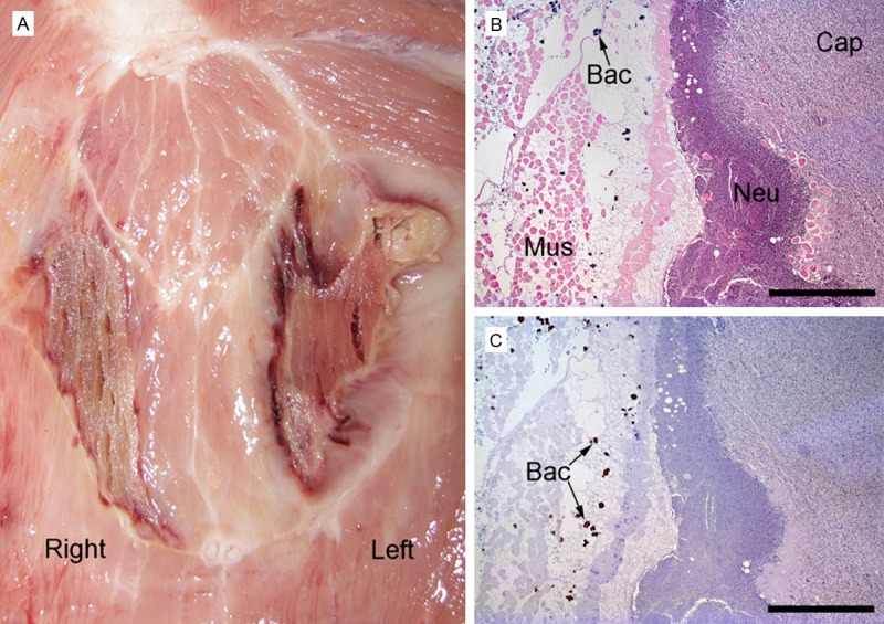
Gross pathology of skeletal muscle necroses in the neck region, pig 1 (A). The lesion in the left muscle has peripheral suppuration. Histopathology of skeletal muscle lesion consisting of necrosis and peripheral suppuration (left-sided lesion in A), (B, C) pig 1. Haematoxylin and eosin stain demonstrates capsule formation (Cap) consisting of granulation tissue, neutrophils (Neu), and necrotic transversely sectioned striated muscle cells (Mus) and bacterial colonies (Bac) (B). Same region stained immunohistocytochemically identifying S. aureus bacteria (Bac) (C). Bar (B, C) = 1 mm.