. 2016 Jan 28;5:e10130. doi: 10.7554/eLife.10130

Figure 5. The kindlin/paxillin complex induces FAK signaling and cell spreading.

(A) FAK and EGFR activation after seeding serum-starved KindCtr, TlnKo and KindKo cells on PLL or FN and treating them with or without EGF and Mn2+. (B) Immunofluorescence staining of activated (Tyr-397 phosphorylated) FAK and F-actin in cells seeded on FN and treated with Mn2+ for 30 min (FAKGFP indicates exogenous expression of FAKGFP fusion protein). (C) FAK and Akt activation in KindKo cells stably transduced with K2GFP or K2ΔPHGFP either seeded on FN or kept in suspension. GFP indicates similar expression of transduced GFP-tagged constructs. GAPDH levels served to control loading. (D) Levels of phosphorylated signaling mediators downstream of FAK in Mn2+-treated, serum-starved or EGF-treated KindCtr, TlnKo and KindKo cells. GAPDH levels served to control loading. (E) Quantification of lamellipodia formation of FN-seeded TlnKo and KindKo cells treated with Mn2+ and either DMSO or the FAK inhibitor PF-228 (n=3 independent repeats; >100 cells/condition; error bars indicate standard error of the mean; significances are given in comparison to the DMSO control). (F) FAK activity in TlnKo and KindKo cells stably transduced with FAKGFP (n=3 independent experiments). (G) Quantification of lamellipodia formation in TlnKo and KindKo cells stably transduced with FAKGFP (n=3 independent experiments; significances are given in comparison to untreated control; error bars indicate standard error of the mean). Bar, 10 µm. DMSO, dimethyl sulfoxide; EGF, epidermal growth factor; EGFR, epidermal growth factor receptor; FAK, focal adhesion kinase; FAKGFP, green fluorescent protein-tagged FAK; FN, fibronectin; GAPDH, glyceraldehyde-3-phosphate dehydrogenase; GFP, green fluorescent protein; PLL, poly-L-lysine.

DOI: http://dx.doi.org/10.7554/eLife.10130.023

Figure 5.

Figure 5—figure supplement 1. FAK phosphorylation in TlnCtr, TlnKo, TlnKo+T1V, KindCtr, KindKo and KindKo+K2GFP cells.

Figure 5—figure supplement 1.

(A) Densitometric quantification of western blot signals of lysates from untreated, EGF- and Mn2+-treated KindCtr, TlnKo and KindKo cells seeded either on FN or PLL and probed with anti-Tyr-397 phosphorylated FAK (pY397-FAK) antibodies (n=3 independent repeats; significances are calculated with respect to PLL adherent cells; error bars indicate standard error of the mean). (B,C) Western blotting of indicated signaling proteins in untreated, EGF- and Mn2+-treated TlnCtr, TlnKo and TlnKo cells re-expressing Venus-tagged talin-1 (TlnKo+T1V) (B), and KindCtr, KindKo and KindKo cells re-expressing GFP-tagged kindlin-2 (KindKo+K2GFP) (C) seeded either on FN or PLL. EGF, epidermal growth factor; FAK, focal adhesion kinase; K2GFP, green fluorescent protein-tagged kindlin-2; FN, fibronectin; GFP, green fluorescent protein; PLL, poly-L-lysine; T1V, Venus-tagged full length talin-1.
Figure 5—figure supplement 2. Kindlin-2 forms a ternary complex with paxillin and FAK.

Figure 5—figure supplement 2.

GFP-IP in lysates of K2GFP-, K2ΔPHGFP- or GFP-reconstituted KindKo cells overexpressing Myc-tagged FAK (FAK-Myc) and Cherry-tagged paxillin (PxnCH). K2GFP but not K2ΔPHGFP forms a ternary complex with paxillin and FAK. FAK, focal adhesion kinase; GFP, green fluorescent protein; IP, immunoprecipitation; K2GFP, green fluorescent protein-tagged kindlin-2.
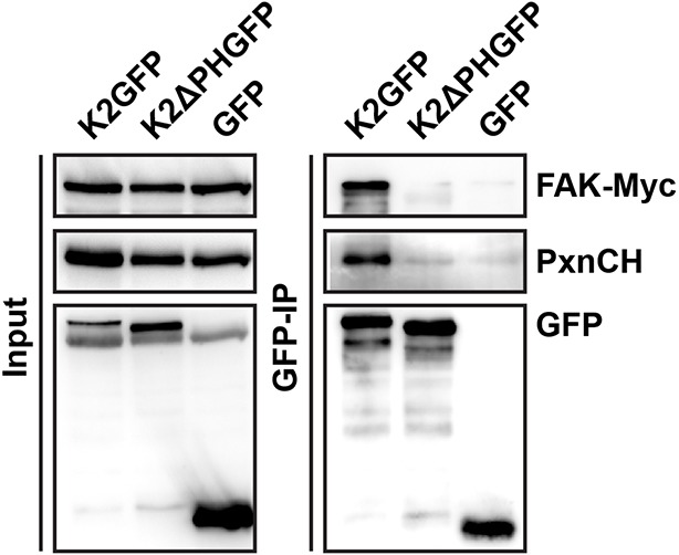
Figure 5—figure supplement 3. Activity of signaling mediators downstream of FAK in TlnCtr, TlnKo, TlnKo+T1V, KindCtr, KindKo and KindKo+K2GFP cells.

Figure 5—figure supplement 3.

(A,B) Western blotting of indicated signaling proteins in untreated (first three lanes), EGF- and Mn2+-treated TlnCtr, TlnKo and TlnKo cells re-expressing Venus-tagged talin-1 (TlnKo+T1V) (A), and KindCtr, KindKo and KindKo cells re-expressing GFP-tagged kindlin-2 (KindKo+K2GFP) (B) seeded on FN. EGF, epidermal growth factor; FN, fibronectin; GAPDH, glyceraldehyde-3-phosphate dehydrogenase; GFP, green fluorescent protein; K2GFP, green fluorescent protein-tagged kindlin-2; T1V, Venus-tagged full length talin-1.
Figure 5—figure supplement 4. Cell spreading of FAK overexpressing TlnKo and KindKo cells.

Figure 5—figure supplement 4.

(A,B) Cell spreading area of TlnKo (A) and KindKo cells (B) seeded on PLL, FN or on FN after FAKGFP overexpression measured by image quantification (n=3; independent repeats are pooled; >100 cells/condition and repeat; resulting areas are shown as binning histograms; significances are calculated between non-transfected cells and FAKGFP expressing cells plated on FN). FAK, focal adhesion kinase; FAKGFP, green fluorescent protein-tagged FAK; FN, fibronectin; GFP, green fluorescent protein; PLL, poly-L-lysine.