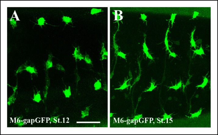
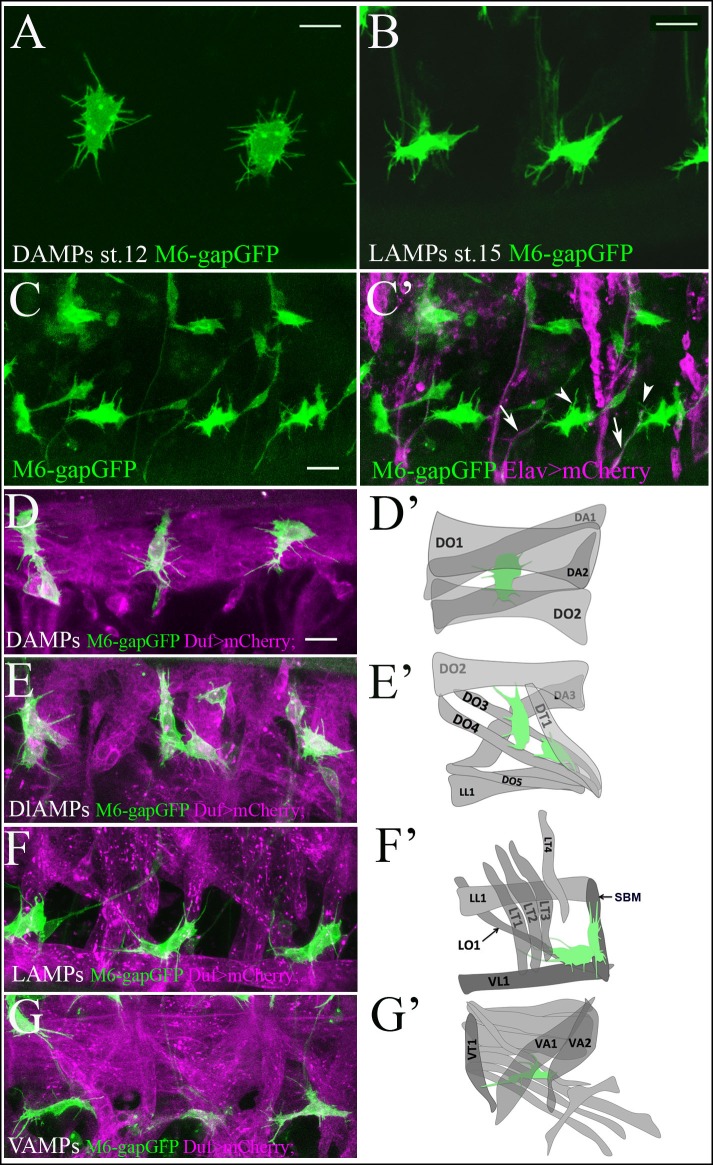
Figure 1. Quiescent AMP cells are tightly associated with surrounding muscles.
(A, B) A zoomed view of quiescent dorsal (A) and lateral (B) AMPs bearing numerous thin filopodia. (A) Newly-specified AMPs at embryonic stage 12 display a random pattern of filopodia. (B) Mid-stage embryo AMPs become elongated and send out filopodia in an directionally-oriented way. Filopodia pattern of AMPs in m6-gapGFP embryos was revealed by anti-GFP staining of membrane-targeted GFP. (C, C’) A lateral view of three hemisegments of stage-15 embryo from the sensor driver line m6-gapGFP; Elav-GAL4; UAS-mCD8mCherry, driving mCherry with a membrane localization signal in all neurons. Arrows point to cytoplasmic extensions connecting the AMPs (green) and aligned with the PNS nerves (magenta). Arrowheads denote thin filopodia that are not connected to the PNS nerves. (D–G) Dual-color in vivo views of three hemisegments of stage-15 embryos from the m6-gapGFP; Duf-GAL4; UAS-mCD8mCherry line. mCherry (magenta) reveals embryonic muscles and GFP (green) reveals AMPs. Dorsal (D), dorsolateral (E), lateral (F) and ventral (G) groups of AMPs are shown. Note that AMPs connect to the embryonic muscles with numerous filopodia. (D’–G’) Schemes represent all observed AMP-muscle connections. AMPs connect to a defined set of muscles. (D’) Dorsal AMP connects to DO1 and DA2 and optionally to DA1 and DO2. (E’) Dorsolateral AMPs connect to DT1, DO3, DO4 and DO2. (F’) Lateral AMPs connect to SBM, LT1, LT2, LT3 and to LO1 and VL1. (G’) Ventral AMP interacts with VA2, VT1 and VA1. Scale bar in (A, B): 4 microns, in (C–G): 9 microns.
Figure 1—figure supplement 1. Segmental pattern of embryonic AMPs.


