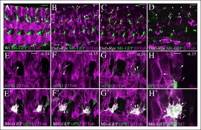
Figure 2. AMP-muscle connections display spatially-restricted plasticity and are decorated by integrin expression.
(A) A wild-type view of AMPs and muscles from mid-stage m6-gapGFP embryo. (B–D) Similar views from m6-gapGFP;Duf-GAL4;UAS-Rpr embryos with (B) weak, (C) intermediate, and (D) strong muscle ablation phenotypes. In segments with partial loss of lateral muscles, the anterior lateral AMP, which normally extends anteriorly (white arrow in A) remained tightly associated with the posterior lateral AMP and interacted mainly with SBM muscle – (white arrows in B and C). In segments with loss of dorsal and dorso-lateral muscles and with some lateral muscles persisting, (B) the dorso-lateral AMPs interacted with remaining lateral muscles (arrowhead in B) to which they do not connect in the wild-type context (arrowhead in A). This indicates a degree of plasticity in AMP connections. In segments with a pronounced loss of dorsolateral and lateral muscles (B and C), the dorsal and dorso-lateral AMPs adopted rounded shapes (yellow arrows) and were unable to migrate to other segments or to the ventral region where muscles were still present. In embryos with total muscle ablation, the majority of remaining AMPs adopted rounded shapes (yellow arrows in D). The number of AMPs detected was drastically reduced (asterisks indicate lacking AMPs). (E–H’) Zoomed views of lateral AMPs stained for (E-G’) α-PS1 and (H, H’) βPS integrin. The first α-PS1 dotty signals associated with AMPs appear at late-stage 14 (E, E’) and are progressively enriched at stages 15 and 16 (F-G’). A punctate α-PS1 pattern is seen, associated with AMP cell bodies (arrowheads) but also aligned with filopodia (arrows in F–G’). A similar β-PS1 pattern denoted by arrows and arrowheads is also observed, starting from embryonic stage 15 (H-H’). Scale bars in (A-D): 30 microns; in (E-G): 10 microns; in (H): 6 microns.

