. 2015 Dec 9;4:e08497. doi: 10.7554/eLife.08497

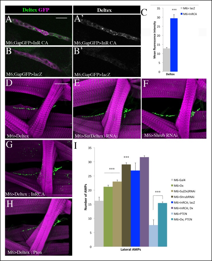
Figure 6. Insulin-driven Notch activation in AMPs involves Deltex.

(A-B’) Single clusters of third-instar larva lateral AMPs stained for Deltex and GFP. (A–A’) There is greater punctate Deltex expression in AMPs expressing constitutively activated InR than in control larva (B-B’) expressing lacZ. (C) Mean fluorescence intensity of the Deltex signal detected in gain-of-function context for Insulin versus wild-type. (D-F) Components of ligand-independent Notch activation have impacts on AMP cell numbers. AMP-targeted expression of Deltex (D), attenuation of Su (Deltex) (E) or attenuation of Shrub (F) all lead to an AMP overproliferation phenotype. The key role of Deltex as an activator of AMP proliferation is confirmed by an increased number of AMPs in embryos with M6-targeted expression of InRCA and Deltex (G) and further supported by partial rescue of AMP number when co-expressing Deltex with the PTEN Insulin pathway inhibitor (H). (I) Graphical representations of mean number of lateral AMPs in genetic contexts shown in (D-H). (***) indicates P ≤ 0.001. Scale bars are (A, B’): 15 microns; (D–H): 45 microns.

DOI: http://dx.doi.org/10.7554/eLife.08497.021

Figure 6—source data 1. Table showing mean number of AMPs in the abdominal segments from the genotypes shown in Figure 6D–I and Figure 6—figure supplement 1 and 2.
For each genotype, the average number of cells ± standard error mean is shown. Sample size (n) is indicated in brackets. Below, a table with mean Deltex signal intensity measurements in lateral AMP cluster from the genotypes shown in Figure 6A–C. For each genotype, the average signal intensity value ± standard error mean is shown. Sample size (n) is indicated in brackets.
DOI: 10.7554/eLife.08497.022

Figure 6.

Figure 6—figure supplement 1. Ligand independent activation of Notch promotes proliferation of AMPs.
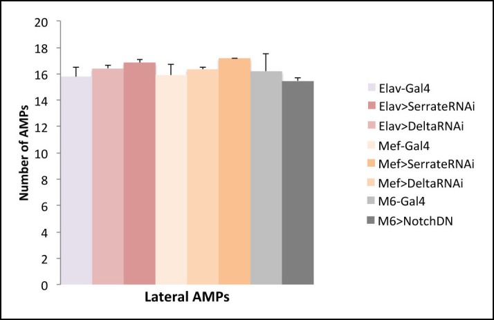
Figure 6—figure supplement 1.

(A) A scheme representing potential sources of Notch ligands (Delta and Serrate). Notch signalling requires cell-cell contact and is activated in AMPs downstream of Insulin. This implies that the activated AMP cell (surrounded by red line) via so far unknown signal X promotes Delta or Serrate expression in cells that are direct AMP neighbours: PNS neurons (in yellow) or muscles (in blue). (B) Notch ligands attenuation in PNS neurons or in muscles and AMP-targeted expression of dominant-negative form of Notch receptor (that binds to ligand but is lacking intracellular domain and thus is unable to activate Notch targets) have no impact on AMP cell number suggesting that Notch activation in AMPs downstream of Insulin receptor is ligand independent.
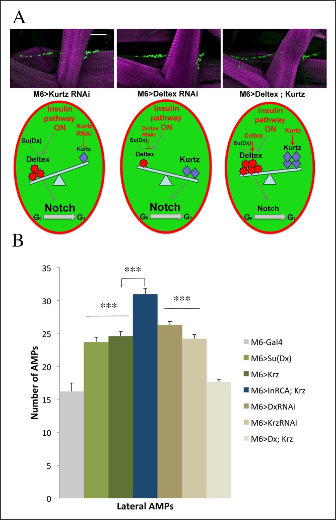
Figure 6—figure supplement 2. Role of Kurtz and Deltex in reactivation of AMPs.

Figure 6—figure supplement 2.

(A) Levels of Deltex versus Kurtz play important role in ligand independent activation of Notch during reactivation of AMPs. Notice that attenuation of Kurtz or Deltex results in an increased number of AMPs whereas simultaneous over-expression of Kurtz and Deltex has no effect on AMP cell number. Schemes below illustrate effects of Deltex and Kurtz levels on Notch activation in an AMP activated by the Insulin pathway. Scale bar: 30 microns. (B) A graph representing the mean number of lateral AMPs of mid third-instar larvae from the genotypes shown in (A) and larvae over expressing in AMPs Su (Deltex), Kurtz and those overexpressing both InrCA and Kurtz. (***) indicates P≤0.001. P-values were computed using t test and Prism software with Gal4 as a control sample except those linked on the graph.