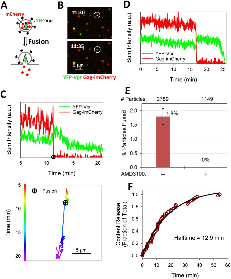
Fig 1. Detection of single HIV-1 fusion based on release of the mCherry viral content marker.
(A) Illustration of viral fusion detection with HXB2 Env pseudotyped particle (HXB2pp) co-labeled with releasable content marker Gag-imCherry (red) and core-associated marker YFP-Vpr (green). (B) HXB2 pseudovirus co-labeled with Gag-imCherry (red) and YFP-Vpr (green) fuses with CV1/CD4/CXCR4 cell as detected by instantaneous loss of mCherry signal. (C) Fluorescence intensity profiles (top) and the virus trajectory (bottom) obtained by tracking the fusing particle in panel B. (D) Fluorescence intensity profiles of another fusing HXB2pp particle. (E) Efficiency of HXB2 Env-mediated single particle fusion in CV1/CD4/CXCR4 cells untreated and treated with 10 μM AMD3100. Total number of particles examined for each condition listed at top of graph. (F) Kinetics of single HXB2pp fusion shown as a cumulative plot of the fraction of fused particles over time. Solid line is obtained by single exponential curve fitting.

