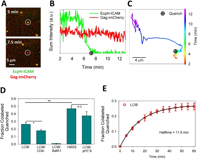
Fig 3. Single particle analyses of HIV-1 entry into acidic endosomes.
(A) HXB2pp co-labeled with EcpH-ICAM and Gag-imCherry enters into a mildly acidic compartment of a CV1/CD4/CXCR4 cell, as detected by quenching of EcpH-ICAM signal. (B, C) Fluorescence intensity profiles and trajectory obtained from tracking the virus particle in panel A show quenching of EcpH signal and subsequent directional movement of the particle. Quenching of EcpH prior to directional movement of the particle is consistent with the initial acidification to pH 6.0 typical for early endosomes. (D) Extent of single-virus particle quenching in CV1/CD4/CXCR4 cells shown as fraction of total particles quenched in one hour experiment for the following conditions: in HBSS, LCIB with or without 100 nM Bafilomycin A1 (BafA1) or LCIB adjusted to pH 7.9. The extent of EcpH-ICAM quenching in parental CV1 cells in LCIB (LCIB/CD4-) is also shown. Error bars are standard error of the mean for three independent experiments. *p<0.05, **p<0.01, “N.S.”p>0.05, student’s t-test. (E) Kinetics of EcpH-ICAM quenching in CV1/CD4/CXCR4 cells bathed in LCIB. Symbols and error bars (red) are mean and standard error of the mean for three independent experiments and curve (black) is fit to single-exponential model.

