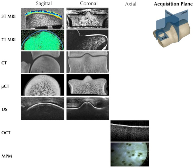
Figure 1.
Each imaging modality can be used to collect information about the tissue, often in multiple acquisition planes. This information can be combined to provide a comprehensive picture of the tissue. Magnetic resonance imaging (MRI) and computed tomography (CT) provide structural information in sagittal, coronal, and axial (not shown) planes, about full thickness cartilage and subchondral bone. Ultrasonography (US) and optical coherence tomography (OCT) provide information about the structural integrity of cartilage with limited penetration into bone. Multiphoton microscopy (MPM) can be used to acquire axial images with submicron resolution, including the identification of individual cells, but with limited depth penetration from the surface to ~200 µm.

