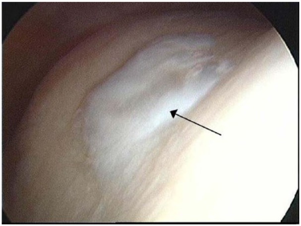
Figure 4.

Representative arthroscopic image of the donor site in a patient 11 months post–cell implantation for autologous chondrocyte implantation (ACI) of the knee. The white portion in the center of the image (black arrow) is the repaired cartilage in the center of the trochlea, observed to be smooth and well integrated into the surrounding cartilage.
