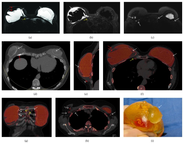
Figure 1.
62-year-old woman presents with enlarging right breast following a fall one month earlier and a history of long standing bilateral subpectoral silicone implants following left modified radical mastectomy and simple right mastectomy in 1992. (a) Axial silicone sensitive MR sequence shows bilateral MR linguini sign of intracapsular rupture (white arrows) with low signal intensity material both within the capsule itself, along with high signal intensity silicone, and within the collapsed right envelope (red arrows). The high signal intensity tissue adjacent to the right internal mammary (IM) vessels (yellow arrow) was not appreciated as being likely due to silicone due to the inhomogeneity of the fat and water suppression about heart. (b) T2 weighted axial IDEAL MR sequence shows high T2 signal fluid of intracapsular seroma within the right envelope (white arrows). There is low signal intensity tissue adjacent to the right IM vessels which in retrospect would be in keeping with silicone within an internal mammary node (yellow arrow). (c) Axial silicone sensitive sequence from the high axilla demonstrates high signal intensity material which was not appreciated prospectively as right intranodal silicone in level I and II nodes and left IM nodes (arrows). (d) Axial mixed energy CT shows the CT linguini sign of collapsed implant envelopes bilaterally (white arrows). Healing fractured right anterior rib is clearly seen (red arrow). (e) Sagittal dual-energy noncontrast CT of the right breast with silicone colored as red shows intracapsular rupture with CT equivalent linguini sign of collapsed silicone envelope (white arrow). Water density material (red arrows) is noted surrounding the collapsed right envelope as seen on fluid sensitive MRI sequence (c). Extracapsular silicone is noted inferiorly (arrowhead). (f) Axial dual energy noncontrast CT of the right breast with silicone colored as red shows bilateral intracapsular rupture with CT equivalent linguini sign of collapsed silicone envelopes (white arrows). Water density material (red arrows) is noted surrounding and within the collapsed right envelope as seen on fluid sensitive MRI sequence (c). Silicone is also easily seen within enlarged right IM lymph node (yellow arrows). (g) Coronal DECT with color coding shows silicone within the right and left internal mammary nodes (white arrows). The internal mammary vessels can be seen (yellow arrows). (h) Axial DECT with silicone colored as red show silicone within right level I and II and left level I nodes (arrows). (i) Photograph of the explanted right implant with extruded silicone from the envelope (white arrow). Serosanguineous fluid noted within the envelope as seen on MRI and CT (red arrow).

