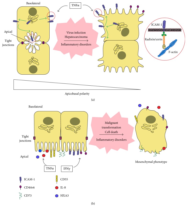
Figure 2.
Redistribution of surface receptors and soluble chemoattractants upon loss of apicobasal polarity in epithelial cells. (a) In polarized hepatocytes, ICAM-1 is able to reach the basolateral membrane but is rapidly redirected to the apical membrane domains. Upon hepatic cell depolarization or in response to persistent stimulation with the inflammatory cytokine TNFα, ICAM-1 and ezrin-radixin-moesin (ERM) proteins, which connect the receptor to the underlying actin cytoskeleton, are oriented towards the stromal milieu and become accessible to immune cells and small hepatic vessels [7]. Other apically polarized receptors involved in leukocyte adhesion, which also interact with the submembranal actin cytoskeleton, such as CD44 isoforms, should be more exposed to parenchymal immune cells upon loss of apicobasal polarity. (b) Columnar epithelial cells, such as intestinal cells, express on their apical membrane key chemokines and lipid mediators, adhesion receptors and other membrane proteins involved in the mobilization of immune cells to the luminal side of the epithelial barrier during inflammation. The proinflammatory cytokine IFNγ is central to increase the expression of some of these receptors, namely ICAM-1 [8]. TNFα may also contribute, for example, by stimulating IL-8 secretion from the basolateral or the apical membrane domains. Following epithelial pathologies or cell death, epithelial cells acquire a “mesenchymal phenotype” and therefore, it is plausible to speculate about the loss of polarized distribution of immune cues also in these epithelial cells. However, the relationship between apicobasal polarity and leukocyte adhesion remains to be investigated in columnar epithelial cells.

