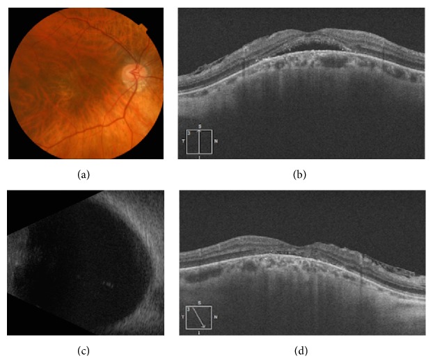
Figure 2.
Staphyloma edge with serous retinal detachment before and after treatment with spironolactone. (a) Color photo showing a tilted-disc with a type 5 staphyloma described by Curtin [2], inferior peripapillary atrophy, marked retinal thinning with visible choroidal vessel, but no hemorrhage in the foveal area. (b) Pretreatment OCT image showing serous retinal detachment in the foveal area with associated epiretinal membrane. (c) B-scan ultrasound showing an abnormal ocular wall in the posterior fundus. (d) Posttreatment OCT image showing resolution of the serous retinal detachment in the foveal area with an abnormal ellipsoid line and associated epiretinal membrane.

