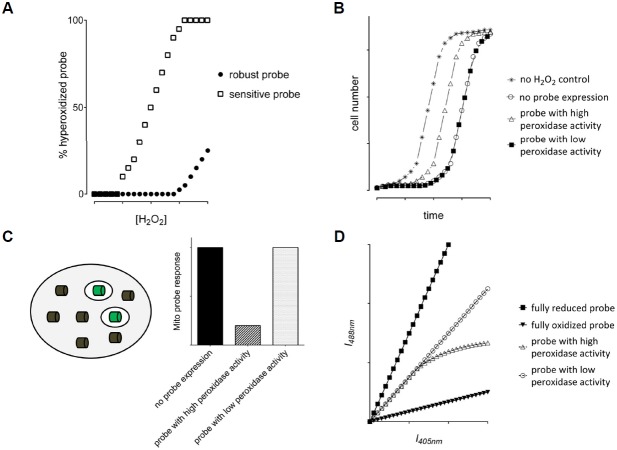
Fig. 3.
Strategies to investigate hyperoxidation sensitivity (A) and physiological impact (B–D) of Prx-based sensors. (A) The fraction of hyper-oxidized probe as a function of the applied H2O2 concentration is determined by hyperoxidation-specific immunoblotting. (B) A growth recovery experiment can be used to determine the influence of the probe expression on cell growth and survival. A theoretical growth curve upon treatment with sublethal concentrations of H2O2 is shown for probes with and without a physiological impact. (C) Investigating the influence of a probe on H2O2 scavenging capacity. A non-fluorescent probe version (grey cylinder) is expressed in the cytosol and its impact on H2O2 homeostasis is measured using a fluorescent probe (green cylinders) expressed in mitochondria. A theoretical result is shown on the right for probes with and without an impact on H2O2 homeostasis. (D) Expected fluorescence intensity scatter plots for a ratiometric probe with low (open circles) and high H2O2 scavenging activity (open triangles).

