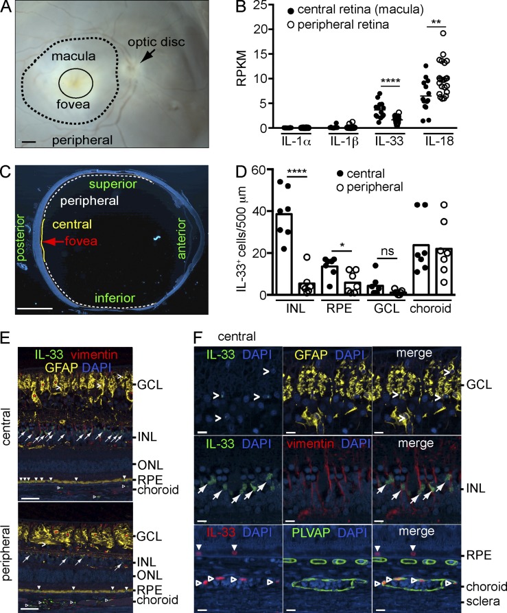
Figure 1.
Expression of IL-33 in Müller cells of the human macula. (A) A representative stereoscope image of a human retina showing the areas of macula and peripheral retina dissected for RNA-sequencing. Bar, 1 mm. (B) RNA-sequencing analysis of IL-1α, IL-1β, IL-18, and IL-33 expression presented as reads per kilobase per million total reads (RPKM) in the macula (n = 14 eyes) and peripheral retina (n = 22 eyes) of control donor eyes. (C) Representative cross section of a human donor eye with central and peripheral areas for quantitative analysis indicated. Bar, 5 mm. (D) Quantification of IL-33+ cells in each retinal layer of central and peripheral areas from seven control donor eyes with age range of 67–89 yr (median age 84, five males and two females). (E) Representative images of immunohistochemical triple staining of IL-33 (green), vimentin (red), and GFAP (yellow) in a control eye from an 84-yr-old male donor. Angle brackets, IL-33+ astrocytes in the GCL; arrows, IL-33+ cells in the INL; filled arrowheads, IL-33+ cells in the RPE; open arrowheads, IL-33+ cells in the choroidal vasculature. Bars, 50 µm. (F) Representative high magnification images of IL-33+ cells in the GCL, INL, RPE, and choroid in the central area of a control donor eye. Shown are IL-33+ astrocytes (GFAP+, angle brackets) and IL-33+ Müller cells (vimentin+, arrows). IL-33+ endothelial cells (open arrowheads) of the choroidal vasculature are shown by IL-33 (red) and PLVAP (green) co-staining. Closed arrowheads, IL-33+ RPE. DAPI (blue), nuclear stain. Bars, 10 µm. GCL, ganglion cell layer; INL, inner nuclear layer; ONL, outer nuclear layer. *, P < 0.05; **, P < 0.01; ****, P < 0.0001; ns, nonsignificant; unpaired two-tailed Student’s t tests.

