Figure 2.
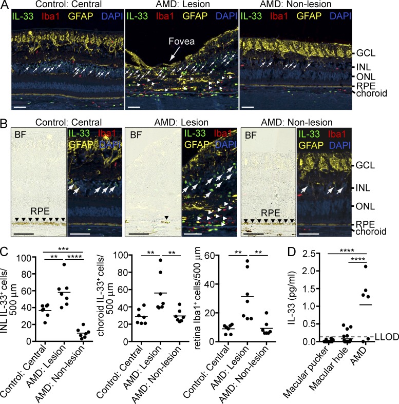
Increased expression of IL-33 in areas of macular degeneration. (A) Immunohistochemical triple staining of IL-33 (green), Iba1 (red), and GFAP (yellow) in the central retina of a control donor eye from an 84-yr-old male with no history of ocular diseases and from an eye of an 82-yr-old female donor diagnosed with AMD. Numbers of IL-33+ Müller cells and mononuclear phagocytic cells (Iba1+ cells) were quantified in areas of retina degeneration (AMD: Lesion), adjacent areas of the same donor that did not exhibit retina degeneration (AMD: Non-lesion), and the central retina of the control donor eye (Control: Central). Arrows, IL-33+ cells in the INL; arrowheads, Iba1+ cells in the subretinal space. DAPI (blue), nuclear stain. Bars, 50 µm. (B) High magnification images representing immunochemistry of IL-33 (green), Iba1 (red), and GFAP (yellow) in the central retina of a control eye and lesion and nonlesion areas of an AMD eye. The bright-field (BF) images show RPE loss in the AMD lesion site. Bars, 50 µm. (C) IL-33+ cells in the INL and the choroid and Iba1+ cells in the retina were counted along an ∼500-µm section within the central retina of seven control donors aged 67–89 yr (median age 84) and lesion and nonlesion areas of eyes from seven AMD donors aged 82–92 yr (median age 86). (D) IL-33 levels in the vitreous from AMD and control patients. IL-33 concentrations in vitreous samples obtained from AMD patients (n = 6, one male and five females, age 68–91, median age 79) and control patients with macular pucker (n = 12, three males and nine females, age 56–79, median age 72) and macular hole (n = 21, 5 males and 16 females, age 46–75, median age 65) were measured by ELISA. **, P < 0.01; ***, P < 0.001; ****, P < 0.0001; one-way ANOVA with Tukey’s post-tests. Horizontal bars represent means.

