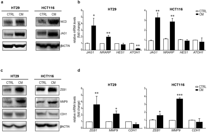
Figure 2. NOTCH1 pathway and EMT markers in HT29 and HCT116 cells under inflammatory conditions.
(a) Western Blot analyses for NICD and JAG1 in HT29 and HCT116 exposed to CM for 12 h. β-ACTIN was used as housekeeping protein. Each experiment was repeated at least three times. (b) qRT-PCR for JAG1 and NRARP, HES1 and ATOH1 in HT29 and HCT116 cells incubated in CM; Statistical significance was calculated on logarithmic transformed values using unpaired t-test, (n = 3, N = 4). *p < 0.05; **p < 0.01; ***p < 0.001. GAPDH was used as housekeeping gene. (c) Western Blot analyses for ZEB1, MMP9, and CDH1 in HT29 and HCT116 exposed to CM for 12 h. β-ACTIN was used as housekeeping protein. Each experiment was repeated at least three times. (d) qRT-PCR for ZEB1, MMP9 and CDH1 in HT29 and HCT116 cells incubated in CM. GAPDH was used as housekeeping gene. Statistical significance was calculated on logarithmic transformed values using unpaired t-test, (n = 3, N = 4 for HT29 and n = 3, N = 3 for HCT116). *p < 0.05; **p < 0.01; ***p < 0.001.

