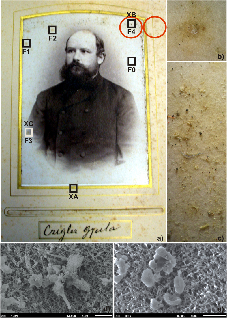
Figure 1. Gyula photograph.
(a) The albumen surface is 9.7 × 13.9 cm; the size of the complete photograph including the cardboard is 10.8 × 16.2 cm. The microbial sampling sites are marked with two red circles. The sites F0–F4 were subjected to FTIR analysis, while the letters XA–XC indicate the sites where XRF measurements were performed. (b,c) Stereo microscope images with a magnification of 40× (site F4) and 20× (area near to site F4) respectively. (d,e) SEM photographs showing the fungal hyphae and spores recovered by a cellulose nitrate membrane. The photograph was supplied by the Slovak National Archive and is used with permission.

