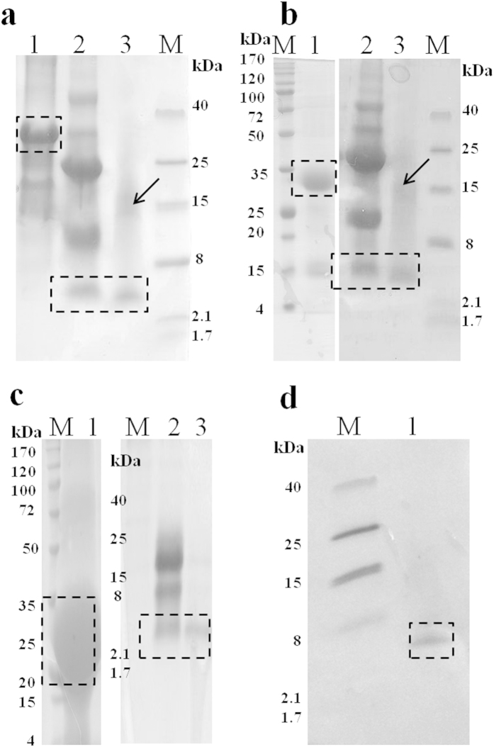
Figure 3. Purification of PG-1, PMAP-36, and Buforin-2 and confirmation of PG-1 expression.
Insoluble protein purification by affinity chromatography, CNBr digestion of affinity-purified target proteins, and reverse-phase high performance liquid chromatography (RP-HPLC) of CNBr-digested r5M-172-PG1-173 (a) r5M-172-PMAP36-173 (b) and r5M-172-Bf2-173 (c) respectively. Lane 1: affinity purification of insoluble protein; lane 2: CNBr digestion of purified insoluble protein; lane 3: final RP-HPLC purification of PG-1 (a) PMAP-36 (b) and Buforin-2 (c) respectively, and lane M is molecular weight marker. The boxes indicate the expected size of the target protein after the various purification steps for PG-1 (2.4 kDa), PMAP-36 (4.2 kDa), and Buforin-2 (2.3 kDa). In lane 3, the purified peptides with higher molecular weights (indicated by an arrow) show the occurrence of AMP multimerization. The samples were loaded onto either 16% Tris-Tricine or 12% SDS-PAGE gels. (d) Confirmation of the presence of purified PG-1 using a rabbit anti-PG-1 antibody.

