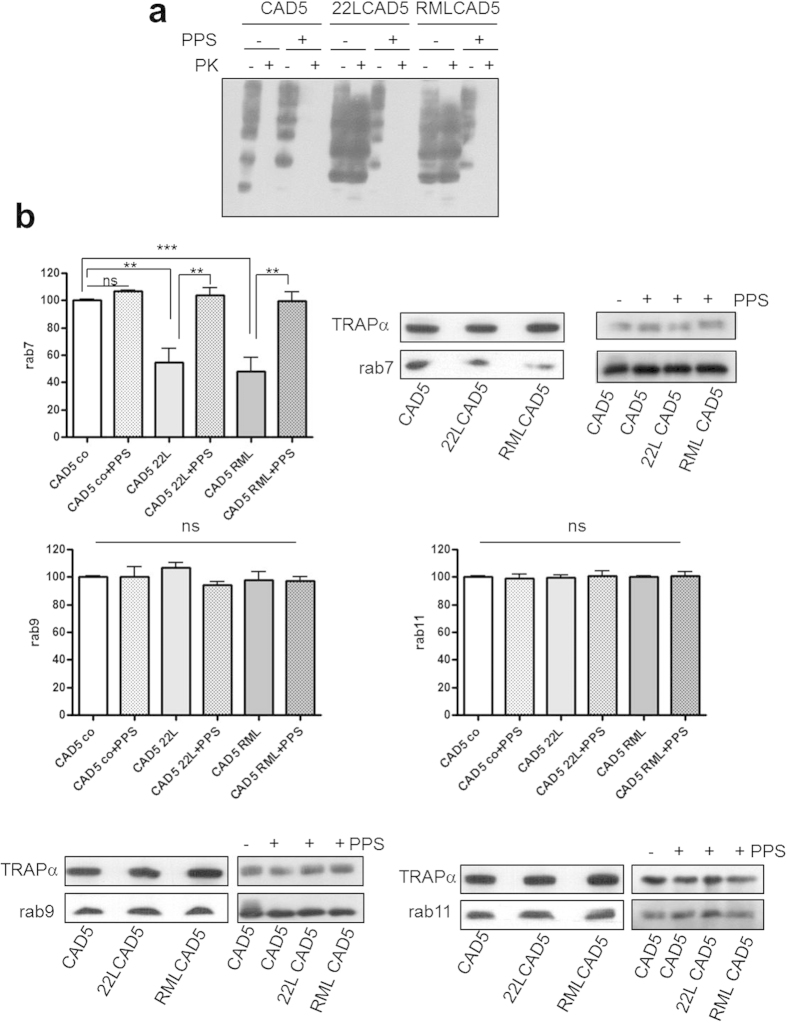
Figure 1. Reduced membrane association of rab7 in CAD5 cells infected with prion strains 22L or RML.
(a) Lysates of CAD5, 22LCAD5 or RMLCAD5 cells −/+ PPS treatment (1 μg/ml for 10 days) as indicated were subjected to PK digestion (20 μg/ml, 30 min, 37 °C) or not. Aliquots were analysed by immunoblot using monoclonal anti-PrP antibody 4H11. (b) Crude membrane preparations (100 μg protein) of CAD5, 22LCAD5 and RMLCAD5 cells −/+PPS as indicated were subjected to SDS-PAGE and levels of rab7, rab9 and rab11, respectively, were analysed by immunoblot. TRAPα served as a loading control. Signals of three independent experiments were quantified by ImageQuant TL (GE Healthcare) and statistical evaluation using one-way ANOVA test, followed by post-hoc analysis with Tukey’s test (GraphPad Prism software; **p-value < 0.01; ***p-value < 0,001; ns = not significant). Bars represent standard deviation.

