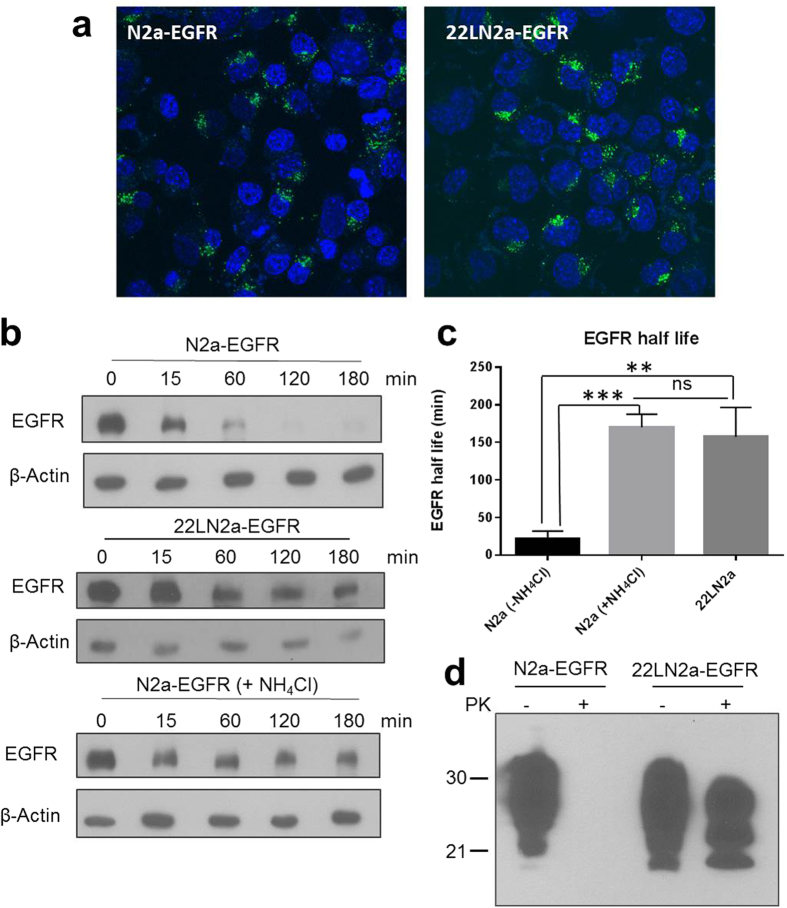
Figure 5. Lysosomal degradation is impaired in 22LN2a cells.
(a) N2a and 22LN2a cells were transduced with recombinant retroviruses encoding human EGFR. Expression of EGFR was confirmed by visualising the internalisation of Alexa488-labeled EGF (green) using confocal microscopy. Nuclei were stained with Hoechst 33342 (blue). (b) EGFR degradation was monitored in N2a-EGFR, 22LN2a-EGFR and N2a-EGFR + NH4Cl cells. Upon pre-treatment with cycloheximide or NH4Cl (if indicated) cells were either directly lysed (0 min), or EGF (50 ng/ml) was supplemented and cells were lysed at indicated time points after EGF addition. Lysates were analysed by immunoblot using an anti-EGFR antibody (upper panel) or anti-β-actin (lower panel) to control for equal loading. (c) The half life of EGFR (signal intensity of 50% compared to the 0 min time point) was determined from three independent experiments upon quantification of EGFR signals and normalization against β-actin signals. Statistical analysis was performed using one-way ANOVA test followed by Tukey’s test (**p-value < 0.01; ***p-value < 0,001; ns = not significant). (d) Representative analysis of PrP signals in lysates of N2a-EGFR and 22LN2a-EGFR with (+PK) or without (−PK) PK digestion.

