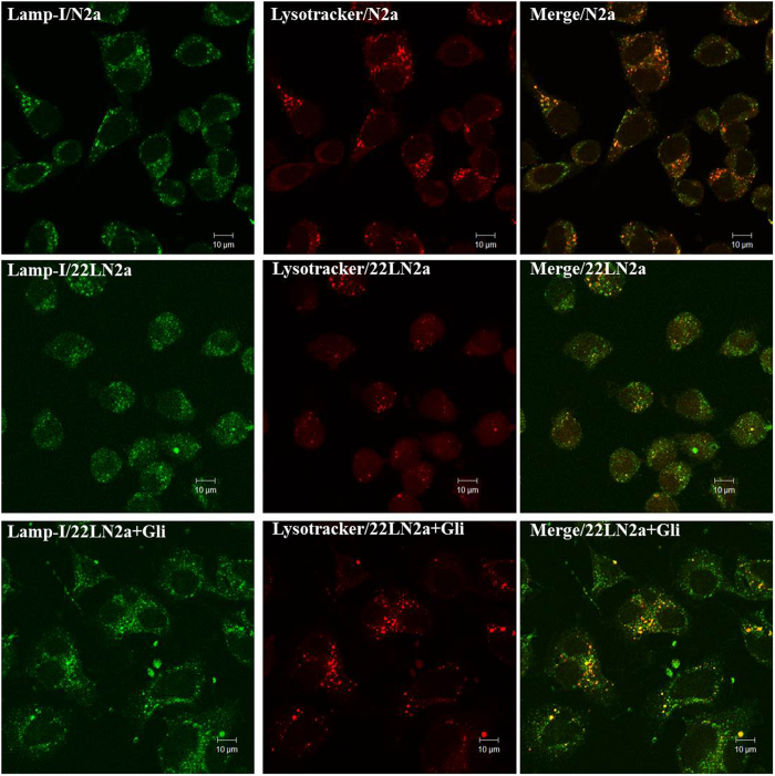
Figure 6. Distribution of late endosomes and lysosomes in N2a and 22LN2a cells.
N2a (upper panel), 22LN2a (middle panel) and 22LNa + Gli (lower panel) were incubated with lysotracker-red (red) for 30 min. Then cells were fixed and stained using an anti-lamp-1primary antibody followed by cy-2-conjugated secondary antibody (green). Yellow colour indicates co-localisation. Confocal images were taken using a Zeiss LSM710.

