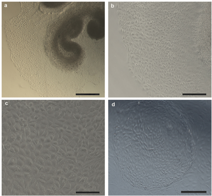
Figure 1. Marginal cells culture under light microscope.
(a) Proliferated marginal cells grew outside the stria vascular explant and were arranged like paving stones with polygonal shape after 3 days of culture (50×), Scale bars, 400 μm. (b) Proliferated marginal cells grew outside the stria vascular explant in 3-day old cultures (100×), Scale bars, 200 μm. Larger magnification is shown in (c) (200×), Scale bars, 100 μm. (d) Proliferated marginal cells were arranged like paving stones, and formed a “cell island” in 3 day-old cultures (100×), Scale bars, 200 μm.

