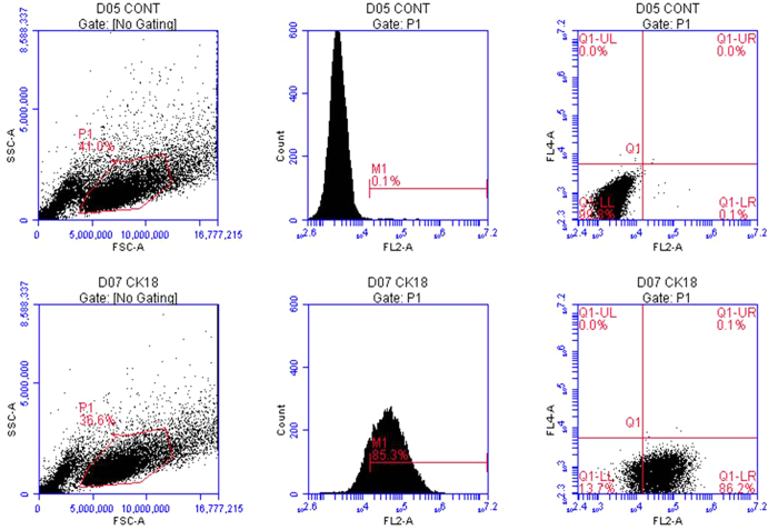
Figure 2. Verification of cultured marginal cells by flow cytometry.
Images in the first row are marginal cells treated with FITC AffiniPure Goat Anti-Mouse IgG (H+L) (negative control). The second row contains marginal cells incubated with anti-cytokeratin 18 IgG and FITC AffiniPure Goat Anti-Mouse IgG (H+L). Flow cytometry confirmed that 85.3% of the cells were cytokeratin 18-positive cells.

