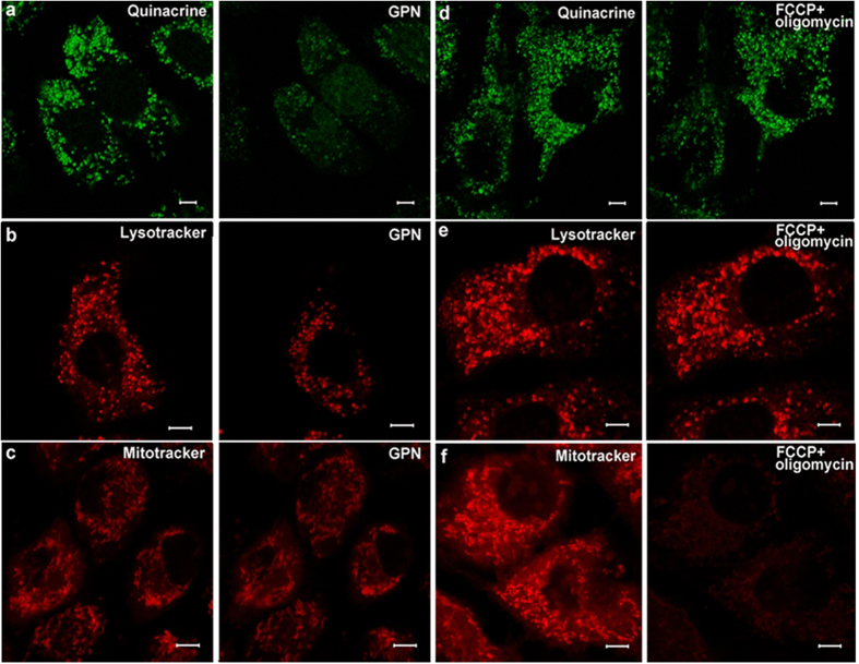
Figure 5. Images of puncta labeled by dyes treated with 200 μM GPN or FCCP (1 μM) + oligomycin (10 μM) for 15 min.
Row (a) Left: green fluorescent punctas in the cytoplasm in a cultured marginal cell incubated with quinacrine; Right: Quinacrine stained puncta was quenched after treatment with 200 μM GPN for 15 min. Row (b) Left: red punctas in a cultured marginal cell after incubation with LysoTracker® Deep Red; Right: LysoTracker stained puncta within the cell was attenuated after treatment with 200 μM GPN for 15 min. Row (c) Left: red punctas revealed mitochondria in cultured marginal cells incubated with MitoTracker® Red CMXRos; Right: Red fluorescence did not change after treatment with 200 μM GPN for 15 min. Row (d) Left: green fluorescent punctas in the cytoplasm in cultured marginal cells incubated with quinacrine; Right: Quinacrine stained puncta did not change in cells after treatment with FCCP(1 μM) + oligomycin (10 μM) for 15 min. Row (e) Left: red punctas in cultured marginal cells after incubation with LysoTracker® Deep Red; Right: LysoTracker stained puncta within the cell did not change after treatment with FCCP(1 μM) + oligomycin (10 μM). Row (f) Left: red punctas revealed mitochondria in cultured marginal cells incubated with MitoTracker® Red CMXRos; Right: red punctas vanished after treatment with FCCP(1 μM) + oligomycin (10 μM).

