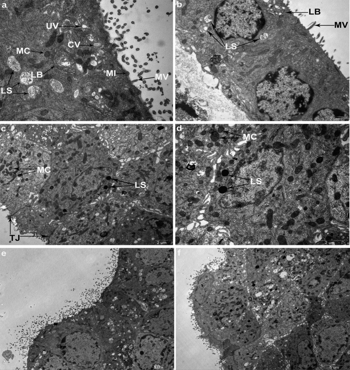
Figure 7. TEM revealed characteristics of marginal cells and lysosomal exocytosis.
(a) TEM photograph shows characteristics of the marginal cell. Arrows indicate lysosomes (LS), mitochondria (MC), coated vesicles (CV), uncoated vesicles (UV), small invaginations (MI), microvilli-like extensions (MV), lamellar bodies (LB), respectively. Scale bars, 1 μm. (b) Marginal cells under TEM. Arrows indicate LS, MV and LB, respectively. Scale bars, 2 μm. (c,d) TEMs of marginal cells loaded with quinacrine for 30 min. Arrows indicate quinacrine labeled lysosomes and unlabeled mitochondria. Neighboring cells were connected with tight junctions. Scale bars, 2 μm. (e,f) Lysosomal exocytosis was observed in marginal cells that contained granules stained with quinacrine. TEM showed the characteristics of the marginal cells and lysosomal exocytosis. A coated omega-shaped invagination was found in the apical plasma membrane of the marginal cells (e). Scale bars, 5 μm.

