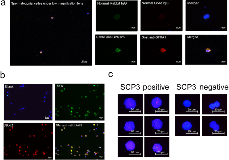
Figure 1. The identification of sorted germ cells.
Germ cells of different differentiated stages were sorted via FACS and MACS, and immune staining were applied to identification the sorted cells. (a) Immunofluorescence identified MACS sorted CD90+ cells. The figure on the left shows cells observed under low magnification lens. The quantity of sorted CD90+ cells was very low. The figures on the right show the detailed staining results. About 90% of these cells were GFRA1 and GPR125 positive, suggested that these cells were enriched undifferentiated spermatogonias. Blank was provided by staining with normal IgGs; (b) Immunofluorescence identified FACS sorted haploid cells. Over 85% of these cells were PRM2 and ACR positive, suggested that these cells were enriched spermatids. Blank was provided by staining with normal IgGs; (c) Spread identified FACS sorted tetraploid cells. Over 200 cells were counted. The figure shows the typical positive cells and negative cells. Over 80% of these cells were definitely SCP3 positive, suggested that these cells were enriched primary spermatocytes.

