
Fig. 1.
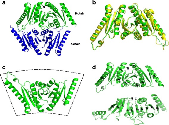
Overall structure of the UspE. a Structure of EcUspE in the tetragonal crystal form, displayed as ribbons. The asymmetric unit contains two protomers colored blue and green. b Structural comparison of the cartoon traces of EcUspE and P. mirabilis USP (PDB code: 4WY2). EcUspE is colored green, and the P. mirabilis USP is colored yellow. The disordered regions are shown with dashed lines. c The monomer structure of EcUspE. d Secondary structural elements of UspE are numbered