
Fig. 1.
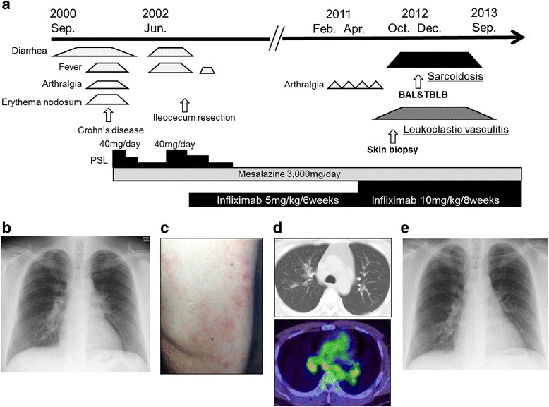
a A chart delineating the clinical course of this case. After infliximab was increased to 10 mg/kg for every 8 weeks, both sarcoidosis and leukocytoclastic vasculitis developed. b A chest X ray on admission revealed bilateral hilar lymphadenopathy. c Scattered eruption on the right thigh on admission. d A Chest CT scan on admission revealed bilateral hilar lymphadenopathy and fine reticulo-nodular shadows in the peri-bronchovascular region of the bilateral upper lobes (upper panel) and positron emission tomography (FDG-PET) scan indicated increased FDG uptakes in the bilateral hilar lymph nodes (lower panel). e Nine months later, the bilateral hilar lymphadenopathy and the upper lobe reticulo-nodular shadows had spontaneously resolved