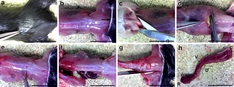
Fig. 2.

Spinal column isolation. a, b After dousing the fur with 70 % ethanol, a small incision is made in the dorsal skin at the level of the hips (a), and the pelt (arrow) removed from the head to hind limbs (b). c The head is removed by cutting at the base of the skull (C1–2 level) and the arms are cut beneath the shoulders to aid removal of the skin. d, e An incision is made through the abdominal wall muscles (d) and continued laterally to the spinal column in both directions (e). f The ribs are then cut parallel with and close to the spinal column on both sides, before detaching the viscera connected to the anterior side of the spinal column. g, h The femurs are cut (g), and the spinal column removed (h) by making a transverse cut at the level of the femurs. In all panels, the head is to the left and tail to the right. The dorsal aspect is imaged in all panels except for d (ventral aspect) and h (C caudal, D dorsal, R rostral, V ventral). Scale bars 2 cm
